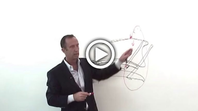
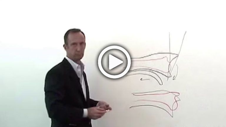
Distal Radius Fractures – Dr. Ruch : Part 3 : Priorities for reduction – Tips
David S. Ruch, MD, Chief of Orthopaedic Hand Service at Duke University Medical Center, provides tips for proper reduction and fixation of intra-articular fractures of the distal radius, including identifying the “critical corner” and establishing where you want your plate to sit on the proximal-distal aspect.
Distal Radius Fractures – Dr. Ruch : Part 5 : Priorities for Reduction – Avoiding Complications
David S. Ruch, MD, Chief of Orthopaedic Hand Service at Duke University Medical Center, discusses the variety of complications that can exist when handling intra-articular fractures of the distal radius, including problems with reduction and over-reduction.
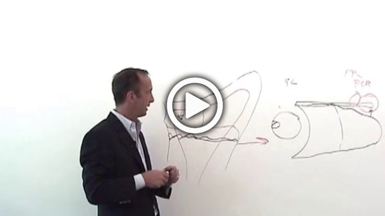
Distal Radius Fractures – Dr. Ruch : Part 6 : Proximal VS standard plate placement
David S. Ruch, MD, Chief of Orthopaedic Hand Service at Duke University Medical Center, discusses the reduction and fixation differences between the Acumed Acu-Loc 2 VDR Proximal Plate and the Acumed Acu-Loc 2 VDR Standard Plate.
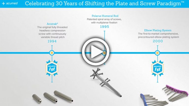
Acumed Innovation Timeline
View a timeline that showcases Acumed’s industry firsts, including Acutrak, Acu-Loc, clavicle plates, and other groundbreaking products.
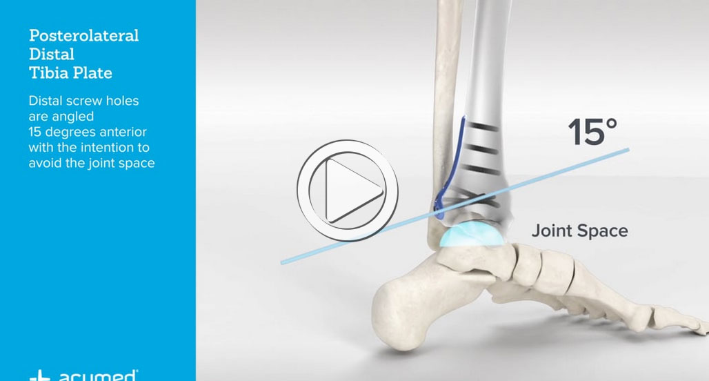
Ankle Plating System 3 Overview
See an overview of the Ankle Plating System 3, a comprehensive system designed to provide a variety of fixation options for fractures of the distal tibia and fibula. Special features include fragment-specific plating for the posterior malleolus, two styles of Hook Plates for avulsion fragments, and a syndesmosis targeting guide. Read More
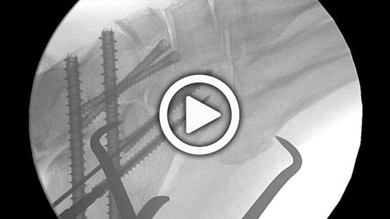
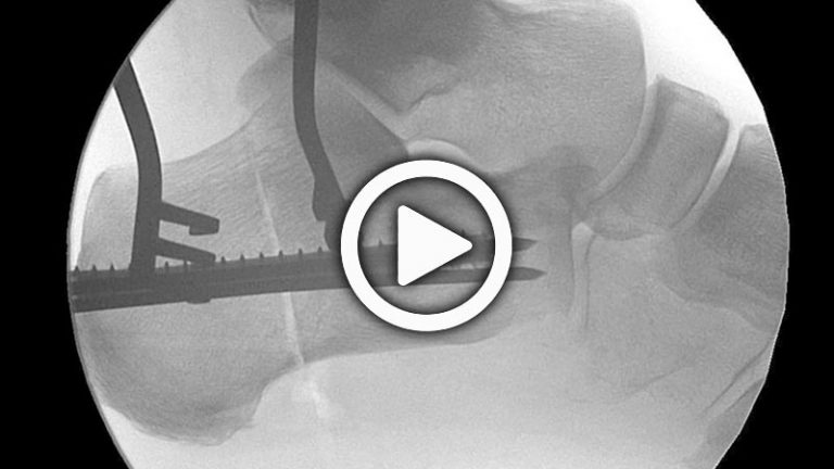
Triple Arthrodesis Cadaveric Lab with Nicholas Abidi, MD
A triple arthodesis fusing the talonavicular, subtalar, and calcaneocuboid joints is performed using Large Acutrak 2 4.7 and 7.5 screws. The rationale for the use of the screws, radiographic evidence, and surgical steps are provided.
Medial Malleolar Fracture Cadaveric Lab with Nicholas Abidi, MD
A medial malleolar fracture is corrected using Large Acutrak 2 4.7 screws. A medial malleolar osteotomy is first performed, and is a component of treatment in osteochondral defects and talar body fractures. The rationale for the use of the screw, radiographic evidence, and surgical steps are provided.
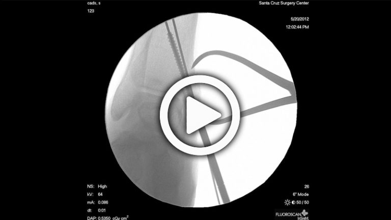
Lateral Malleolar Fracture Cadaveric Lab with Nicholas Abidi, MD
A Weber Type A lateral malleolar fracture is corrected using a Large Acutrak 2 5.5 screw. The rationale for the use of the screw, radiographic evidence, and surgical steps are provided.
Calcaneal Osteotomy Cadaveric Lab with Nicholas Abidi, MD
A medial calcaneal osteotomy is performed to correct an adult flatfoot deformity using Large Acutrak 2 7.5 screws. This procedure could also be done to correct a pes cavus deformity. The rationale for the use of the screw, radiographic evidence, and surgical steps are provided.
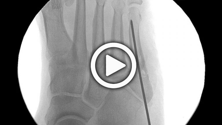
Jones Fracture Cadaveric Lab with Nicholas Abidi, MD
A Jones fracture (5th metatarsal fracture) is corrected using a Large Acutrak 2 5.5 screw. The rationale for the use of the screw, radiographic evidence, and surgical steps are provided.
